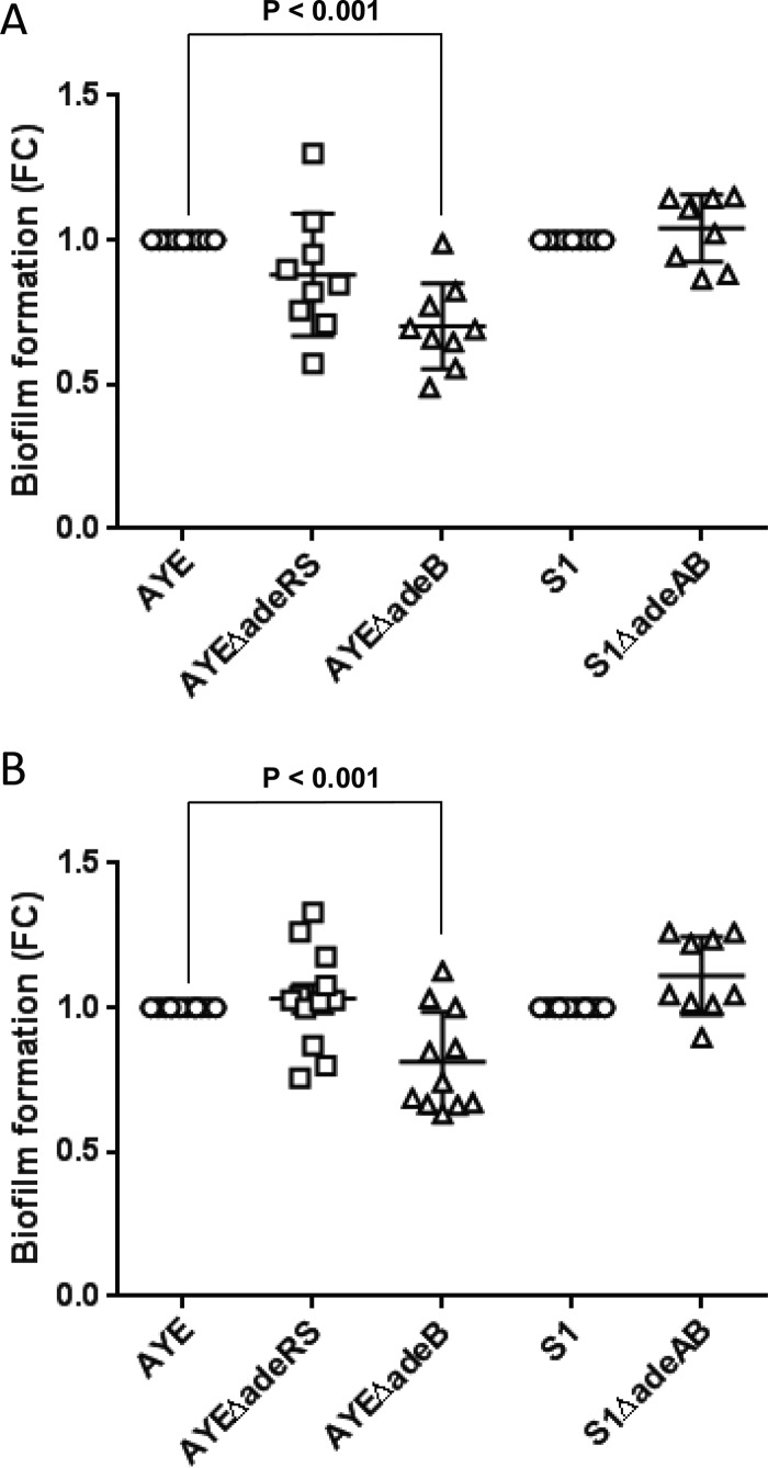
FIG 3 .
Biofilm formation on plastic pegs at 30°C and 37°C as determined by crystal violet staining. Panels: A, 30°C; B, 37°C. Markers show fold change (FC) in OD600 compared with the parental strain in individual biological replicates, with horizontal lines representing the mean and vertical lines and whiskers showing the standard error of the mean.

