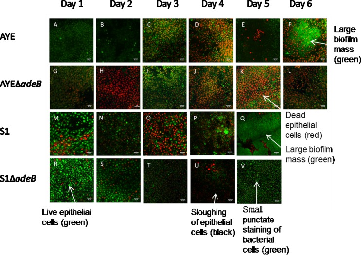
FIG 6 .
Time course of biofilm development on mucosa observed by LIVE/DEAD staining and confocal laser microscopy. Uninfected epithelia are live (green) and intact throughout. Red, rounded epithelial cells indicate epithelial cell death. Small, punctate green staining indicates bacterial cells, and large, green-staining masses indicate bacterial biofilm. Black areas depict the exposed extracellular matrix. Arrows indicate examples of live and dead epithelial cells, bacterial cells, epithelial cell sloughing, and biofilm masses. (A to F) Growth of parental strain AYE on mucosal tissue. (G to L) Growth of strain AYEΔadeB on mucosal tissue. (M to Q) Growth of parental strain S1 on mucosal tissue. (R to V) Growth of strain S1ΔadeAB on mucosal tissue. Images are representative of at least three repeated experiments.

