
FIGURE 2.
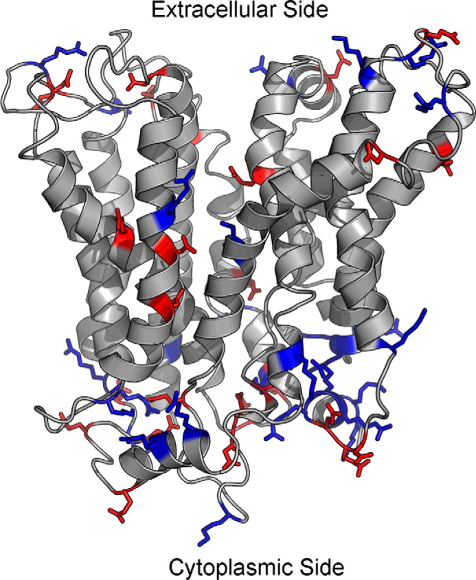
Distribution of positive and negative charges in the outward-partially occluded model of MFSD2A. Majority of the charged residues are located at the top and bottom surfaces of the protein, where there is minimal contact with the hydrophobic lipid bilayer, and maximal contact with the hydrophilic extracellular or intracellular environment. There are more positively charged residues on the cytoplasmic surface of MFSD2A. Blue, positively charged residues; red, negatively charged residues.