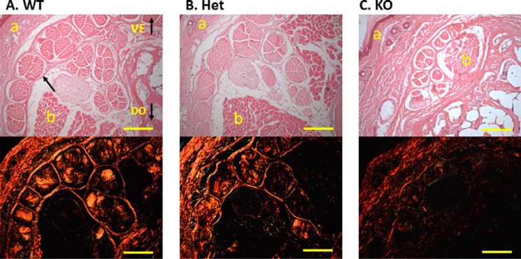
FIGURE 1.
Histological evaluation of tail tendon. Tail tendons were transected and stained with H&E (top) and picrosirius red (bottom) from wild-type (A), heterozygous (B), and CypB KO (C) mice. CypB KO mouse tendon contained fewer collagen bundles, each consisting of fewer fascicles that are poorly organized and have an abnormal morphology. The collagen fibers in the CypB KO tendon showed a green and, in some areas, orange/red color, whereas those of wild-type and heterozygous mouse tendon show a relatively uniform orange color with picrosirius staining. The arrows indicate the single fascicle in the tendon. VE, ventral side of tendon; DO, dorsal side of tendon; a, dermis; b, muscle. Bar, 200 μm.

