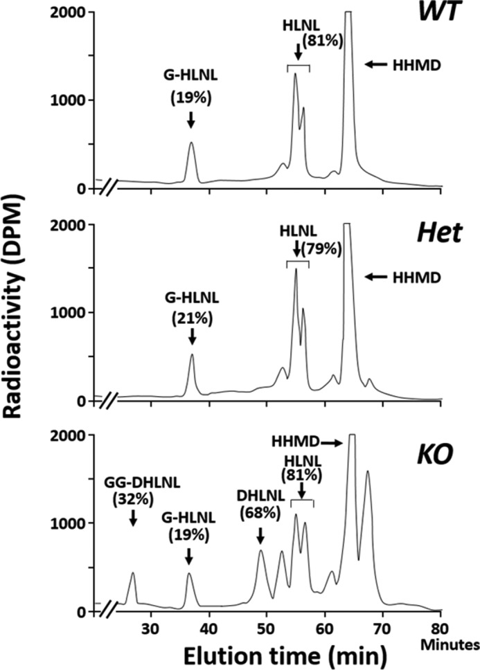
FIGURE 7.

Typical chromatographic patterns of collagen cross-links of the base hydrolysates. Shown are WT (top), Het (middle), and CypB KO (bottom) mice. The amounts of GG-, G-, and free DHLNL and HLNL are shown in percentages (GG-DHLNL + DHLNL = 100%, G-HLNL + HLNL = 100%). In CypB KO tendon, the relative amounts of glycosylated (G-) and non-glycosylated HLNL were comparable with WT and Het tendon. The DHLNL in CypB KO tendon was found to be the diglycosylated or non-glycosylated form. No glycosylation was detected for HHMD.
