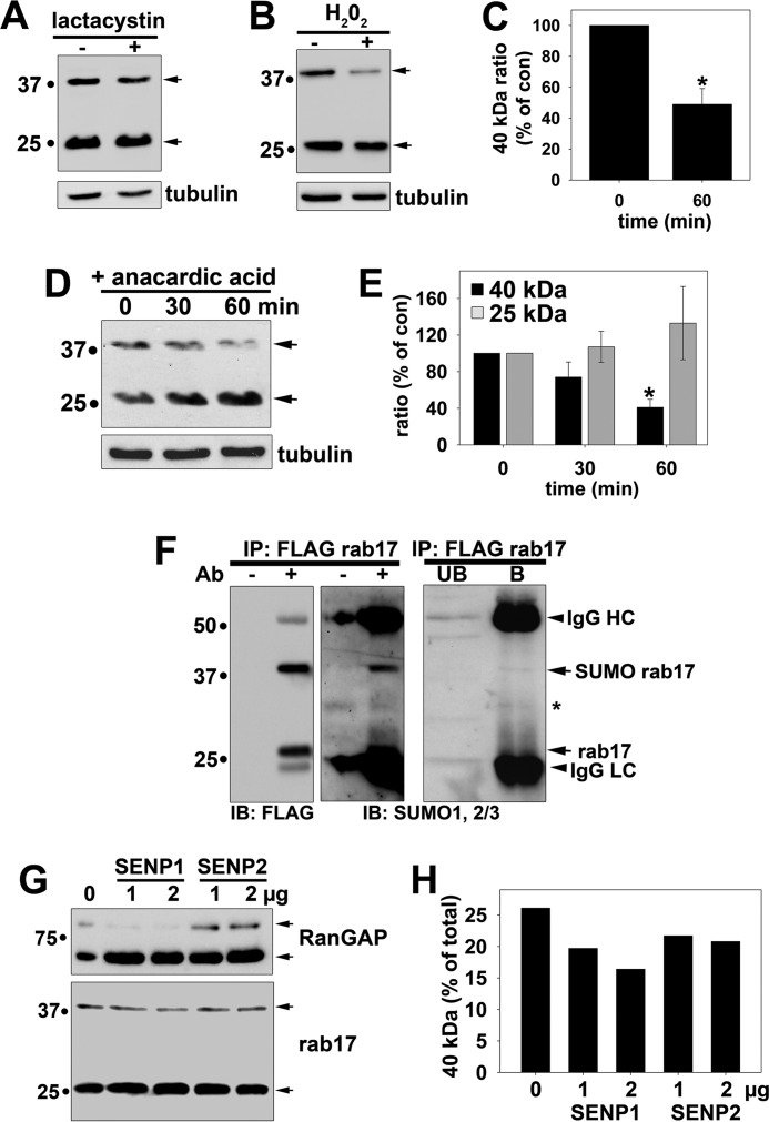
FIGURE 2.
Rab17 is sumoylated. A, Clone 9 cells expressing myc-tagged wild type rab17 were treated with 5 μm lactacystin for 4 h at 37 °C. Lysates were collected and immunoblotted (IB) for rab17 with anti-rab17 antibodies or α-tubulin as a control. B, Clone 9 cells expressing FLAG-tagged wild type rab17 were treated with 1 mm H2O2 for 1 h at 37 °C. Lysates were collected and immunoblotted for rab17 using FLAG antibodies or for α-tubulin as a control. C, the percent of rab17 relative to total was determined by densitometry and plotted as the percent of the control ratio. The values represent the average from at least three independent experiments ± S.E. *, p ≤ 0.002. D, Clone 9 cells expressing myc-tagged wild type rab17 were treated for the indicated times with 5 μm anacardic acid at 37 °C. Lysates were collected and immunoblotted for rab17 with anti-rab17 antibodies or α-tubulin as a control. In E, the percent of 40- or 25-kDa rab17 relative to total was determined by densitometry for each sample and plotted as the percent of the control ratio. The values represent the average from at least three independent experiments ± S.E. *, p ≤ 0.003. F, wild type rab17 was immunoprecipitated from cell lysates with anti-FLAG antibodies and immunolabeled for rab17 (with anti-FLAG antibodies) or antibodies against SUMO1 and SUMO2/3. Lysates without added antibodies (−Ab) served as negative controls for nonspecific protein binding to the Sepharose (middle panels). A lighter exposure of the SUMO immunoblots are shown (right panel). Arrows are marking the cross-reactive IgG heavy (HC) and light chains (LC), the SUMO-positive 40-kDa rab17, and unmodified 25-kDa rab17. The asterisk is marking a cross-reactive species in both bound and unbound samples. Molecular mass markers are indicated on the left in kDa. G, cleared lysates were treated with the indicated concentrations of either SENP1 or SENP2 proteases for 1 h at 37 °C. Reactions were stopped by addition of Laemmli sample buffer and immunoblotted for RanGAP (positive control; upper panel) or rab17 (lower panel). Arrows indicate the sumoylated (top-most arrow for each panel) and desumoylated (bottom-most arrow) forms of the proteins. A representative immunoblot from at least three independent experiments is shown. In H, the levels of the 40-kDa band relative to total rab17 from the immunoblot in G were determined by densitometry and plotted.

