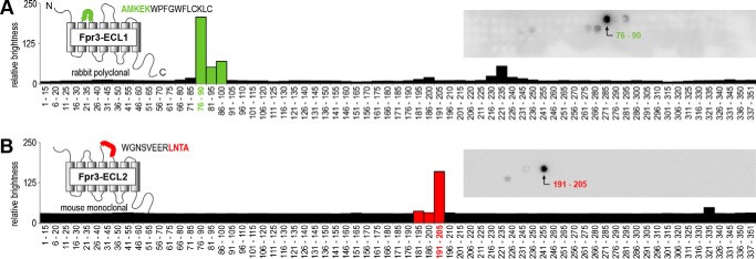
FIGURE 1.
Generation of two novel specific Fpr3 antibodies. Peptide spot array analyses of the polyclonal rabbit antibody Fpr3-ECL1 (A, green) and the monoclonal mouse antibody Fpr3-ECL2 (B, red) are shown. Models indicate the positions and sequences of the immunization peptides used for antibody generation. The amino acid motifs that are recognized by the antibodies are indicated in the respective colors. Both antibodies were tested against peptides comprising the complete Fpr3 sequence. Each spot consists of a 15-amino acid peptide overlapping by 10 residues with its predecessor. Insets show original arrays visualized by enhanced chemiluminescence. The charts show the quantification of staining intensities; bars containing the AMKEK or LNTA motif are highlighted in green or red, respectively. Numbers denote the peptide positions in the receptor protein.

