FIGURE 2.

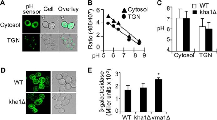
No significant change in the functions of the secretory pathway in Kha1p-dificient cells. A–C, measurement of pH of the Golgi vesicles and cytosol. Cells were transformed with a plasmid expressing a pH-sensitive fluorescent protein at the cytosol or at the lumen of the TGN. A, visualization of subcellular distribution of the pH sensors by fluorescent microscopy. B, a calibration curve reflecting the ratio of emission intensity at the indicated pH. Data represent the average of cells in A600 = 1. C, pH at the cytosolic or lumen of the TGN was determined by using the calibration curve. D, no difference in subcellular distribution of Fet3p fused with YFP (Fet3-YFP) in WT control and kha1Δ cells. E, KHA1 gene knock-out did not lead to unfolded protein response. WT, kha1Δ, and vma1Δ cells were transformed with a reporter plasmid of unfolded protein response. Cells cultured in SC media were subjected to β-galactosidase assays. Data represent the average ± S.D. (n = 4). The asterisk (*) indicates p < 0.05 compared with WT control cells.
