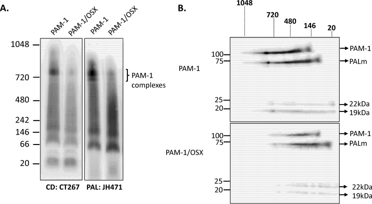
FIGURE 10.
Blue native-PAGE analysis reveals diminished ability of PAM-1/OSX to form complexes in AtT-20 cells. A, solubilized membrane proteins prepared from PAM-1 and PAM-1/OSX AtT-20 cells were separated by blue native-PAGE. Molecular weight markers are indicated to the left. After transfer to a PVDF membrane under native conditions, duplicate blots were probed with antibodies to PAL or PAM-CD. Multiple high molecular weight complexes containing PAM were apparent. B, single gel lanes from PAM-1 or PAM-1/OSX AtT-20 cells were excised, reduced, and denatured and then fractionated by SDS-PAGE. After transfer to a PVDF membrane, PAM proteins were visualized using a CD antibody; PAM products are indicated on the right and native molecular weight markers are shown above the blots. This experiment was repeated three times with separate lysates, with similar results.

