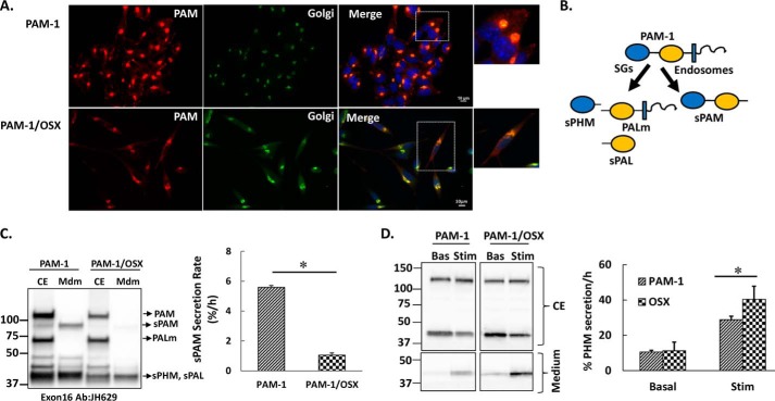
FIGURE 5.
Secretion of PAM-1 products is altered in PAM-1/OSX cells. A, steady state localization of PAM was assessed in AtT-20 lines expressing PAM-1 or PAM-1/OSX; PAM was visualized using an antibody to its cytosolic domain (6E6; Cy3 anti-mouse), and the Golgi complex was visualized using an antibody to TGN38 (FITC anti-rabbit). Scale bar, 10 μm. B, schematic illustrating soluble products of secretory granule (SG) and endosomal processing of PAM-1. C, PAM-1 and PAM-1/OSX cells were incubated for 1 h in basal medium; cell extracts (CE) and media (Mdm) were subjected to Western blot analysis using an antibody to exon 16 (JH629). The basal secretion rate of sPAM, which is produced only in the endocytic pathway, was calculated from these Western blots (% = 100 × sPAMmdm/PAM-1cell) and was diminished in PAM-1/OSX cells (*, p < 0.05, n = 6). D, duplicate wells of AtT-20 cells expressing PAM-1 or PAM-1/OSX were either incubated in basal medium (Bas) or medium containing 2 mm BaCl2 (Stim) for 30 min; cell extracts (CE, upper) and spent medium (lower) were subjected to Western blot analysis using an antibody specific to PHM (JH1761). Secretion rates (% cell content/h) were calculated by taking the ratio of sPHM in the medium to sPHM in the cell extract, although its basal secretion did not differ, the stimulated secretion of sPHM was increased in PAM-1/OSX cells (*, p < 0.05, n = 3).

