FIGURE 6.
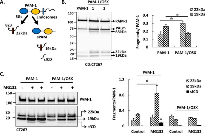
Endocytic processing of PAM-1/OSX is altered. A, schematic shows membrane-associated C-terminal fragments of secretory granule (SG) and endosomal processing of PAM-1 in AtT-20 cells. B, samples from Fig. 4A were analyzed using an affinity-purified antibody specific for the C terminus of PAM (CT267). Signals for 19- and 22-kDa intermediates in the two cell types from two independent experiments were quantified and expressed as a percentage of intact 120-kDa PAM-1 (*, p < 0.05, n = 6). C, PAM-1 and PAM-1/OSX cells (duplicate wells) were incubated in basal medium without (−) or with (+) MG-132 for 4.5 h. Cell lysates were analyzed using the PAM-CD antibody. Signals for sfCD, 19- and 22-kDa intermediates in MG-132-treated versus control wells from two independent experiments were quantified and expressed as a percentage of intact 120-kDa PAM-1 (* for the effect of MG-132, p < 0.05, n = 6).

