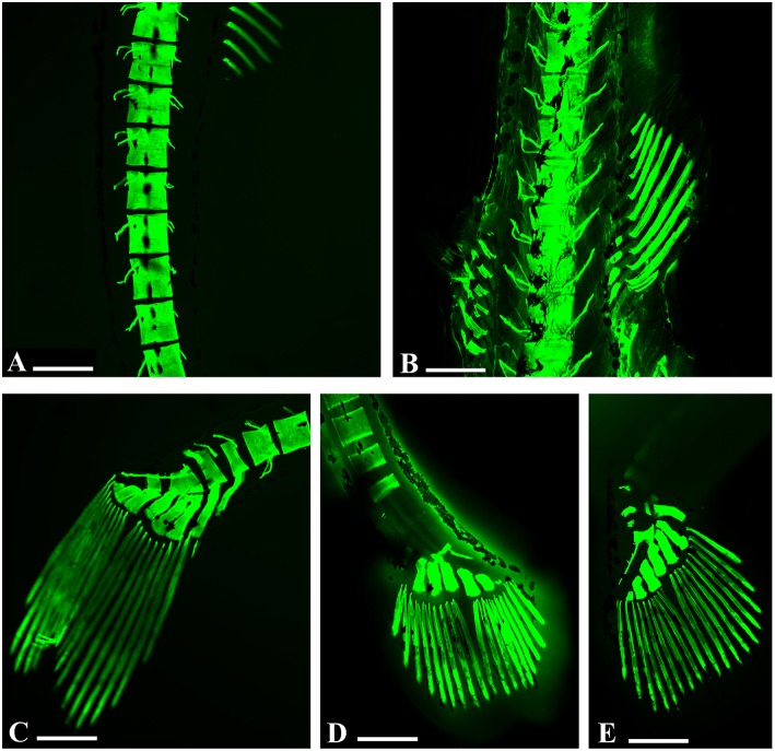
Figure 3.
Embryo zebrafish treated with calcein solution. (A,B) Particularly abnormalities in vertebral spines in samples treated with 200 mg/L. (A) Zebrafish embryos 16 days. (B) Zebrafish embryos 21 dpf. (C–E) Zebrafish embryos 16 days. Details of the abnormalities of the caudal rays with clear areas of decalcification of caudal vertebrae related to the concentrations tested. (C) 50 mg/L; (D) 100 mg/L; (E) 200 mg/L. Scale bar: 200 μm.

