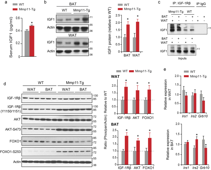
Figure 4. MMP11 activates the IGF1/AKT/FOXO1 axis.
(a) Circulating IGF1 levels in Mmp11-Tg mice and controls (n = 8/group); (b) Immunoblots describing the expression of IGF1 in the WAT and BAT of Mmp11-Tg mice and controls; (c) Increased physical interaction between IGF1 and its receptor IGF1Rβ in the WAT and BAT of Mmp11-Tg compared to WT mice. Protein extracts were immunoprecipitated with an IGF1Rβ antibody and the interaction was revealed with an anti-IGF1 antibody; (d) left panel: Immunoblots for proteins involved in the IGF1 signalling pathway in the WAT and BAT of Mmp11-Tg mice compared to WT, right panel: quantification of the ratios of phosphorylated IGF1Rβ, pAKT, and pFOXO1 proteins relative to IGF1Rβ, AKT, and FOXO1, respectively, normalized to actin expression (data are presented as fold change of Mmp11-Tg values to WT values); (e) Expression profile of genes involved in the insulin/IGF1 pathway in the WAT and BAT of Mmp11-Tg and WT mice (n = 6–7/group).

