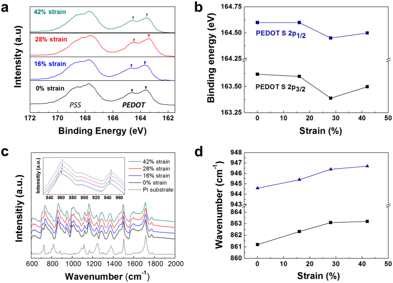
Figure 3. Spectroscopic analysis on the change in bonding energy with tensile strain.
(a) XPS spectra of S 2p for PEDOT:PSS films as a function of applied strain (0, 16, 28, and 42%). (b)The change in binding energy of S 2p in PEDOT according to the strain. (c) FT-IR absorption spectra for PEDOT:PSS films with the tensile strain and PI substrates. The inset shows the peak shift of C-S bonding. (d) The change in carbon-sulfur (C-S) bonding energy in PEDOT chain as a function of the strain.

