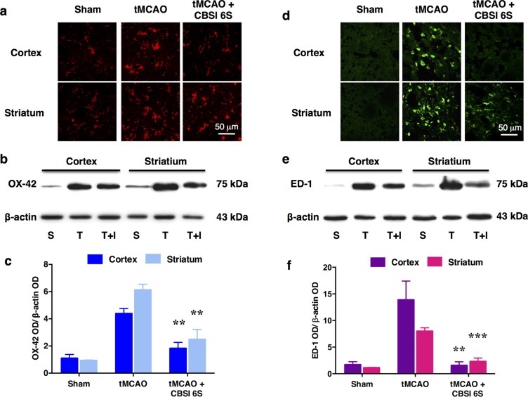
Figure 6.
Reduction of microglial number and activation of the MCAO-affected cortex (Co) and striatum (St) by CBS inhibitor 6S. (a) OX-42-immunopositive microglial cells observed in the Sham-operated (S) group are resting microglia based on their ramified star-like morphology. Marked increase in the number of microglia was observed in the tMCAO (T) group. Many of these cells are activated as they appeared to be large with thick processes. In the CBSI 6S-treated tMCAO (T + I) group, the number of activated microglia was reduced although still increased when compared to the Sham group. (b) Representative Western blot results. (c) Quantified Western blot results, n = 3–4. ANOVA: F (2, 8) = 19.399 for cortical OX-42, F (2, 8) = 25.288 for striatal OX-42, **p < 0.01 against respective tMCAO (T) group by Bonferroni. Western blot results support the observations in A. (d–f) are similar to (a–c) except that they are for ED-1 which is a marker of macrophages including phagocytic microglia, which are comparatively absent in the sham group. (f) ANOVA: F (2, 8) = 15.408 for cortical ED-1 and F (2, 7) = 59.718 for striatal ED-1, **p < 0.01, ***p < 0.001 against respective tMCAO (T) group by Bonferroni.

