
Figure 1. Explanted Renal Allograft Showing Evidence of Rejection and Expression of PD-1 Pathway Molecules after Administration of PD-1 Antibody.
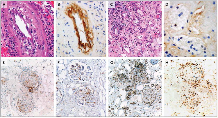
In Panel A (hematoxylin and eosin), arteries show intimal arteritis and focal intimal foam cells, findings consistent with chronic vasculopathy in the allograft. In Panel B, strong immunostaining for C4d (a product of the classical complement pathway) is present in the artery endothelium. Panel C (hematoxylin and eosin) shows glomerulitis, severe tubular loss, tubulitis, interstitial edema, and interstitial inflammation. In Panel D, peritubular capillaries show capillaritis but negative C4d immunostaining. In Panels E and F, immunostaining for programmed death 1 ligand 1 (PD-L1) and programmed death 1 ligand 2 (PD-L2), respectively, show that these molecules are present on endothelial cells and infiltrating immune cells associated with glomeruli. In Panel G, infiltrating T cells expressing programmed death 1 (PD-1) are associated with cells expressing PD-L1 and PD-L2. In Panel H, the infiltrating immune cells are predominantly CD8-positive and are coexpressed with Ki-67, findings consistent with an activated cytotoxic T-cell phenotype; shown are the results of double immunostaining for CD8 (brown chromagen) and Ki-67 (blue chromagen).