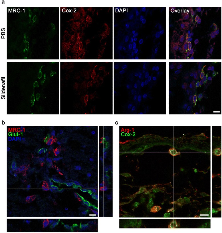
Fig. 7.

3D confocal analysis of double-labeled microglia and vessels after pMCAo and PBS and/or sildenafil (0.5 mg/kg) treatment. a Co-localization of M1 (COX-2 in red) and M2 (MRC-1 in green) markers in the leptomeningeal membranes, 72 h after ischemia. b MRC-1 (in red) is present in the perivascular microglia (in green). c Co-localization of M1 (COX-2 in green) and M2 (Arg-1 in red) markers in the leptomeningeal membranes 8 days after pMCAo, in a PBS-treated animal. Scale bar represents 10 μm (a–c)
