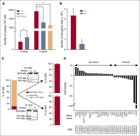
Figure 3.
ABL1 inhibits proliferation of BCR-ABL1 leukemia cells. (A) Mean number of colonies ± SD from BCR-ABL1 Abl1−/−, BCR-ABL1 Abl1+/+, and BCR-ABL1/YFP-ABL1 Abl1−/− [−/−(+)] leukemia cells; *P < .001, **P < .05. (B) Mean number of total clonogenic cells ± SD generated by 106 BCR-ABL1 Abl1−/− and BCR-ABL1 Abl1+/+ cells during 5 weeks in vitro culture; *P < .001. (C) The composition of initial cell mixture (left bar) and those after 5 weeks of in vitro and in vivo expansion (right bars). Western blot analyses using anti-ABL1 and anti-GFP/YFP antibodies (top and bottom boxes in each panel, respectively) show expression of BCR-ABL1 as well as YFP-ABL1 protein in the initial cell mixture (0) and after 5 weeks (5). (D) Statistically significant (FDR < 0.05) fold changes (>1.5) of expression of indicated genes regulating cell proliferation in BCR-ABL1 Abl1−/− vs BCR-ABL1 Abl1+/+ leukemia cells maintained with SCF + IL-3.

