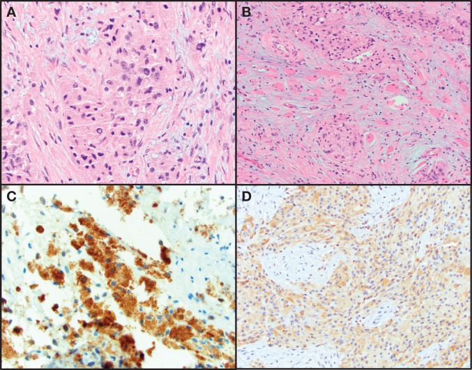
Figure 1.
Hematoxylin and eosin (H&E)-stained images of a granular cell tumor. (A) High magnification of pleomorphic polygonal cells with granular cytoplasm. (B) Granular cell neoplasm infiltrating skeletal muscle. (C) Positive immunohistochemical staining for S100. (D) Positive immunohistochemical staining for CD68.

