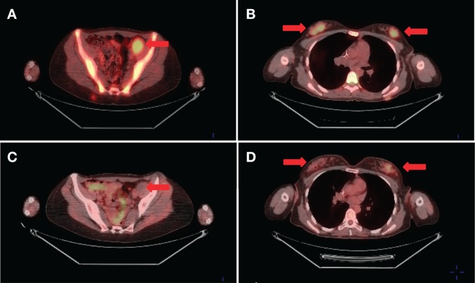
Figure 2.
Representative fused FDG-PET images prior to (A,B) and 3 mo after (C,D) starting pazopanib. SUVmax at the left pelvic metastatic lesion was 5.0 prepazopanib (A) and not hypermetabolic at 3 mo (C). SUVmax in the right breast metastatic nodules was 5.8 prepazopanib (B) and 2.7 after treatment (D).

