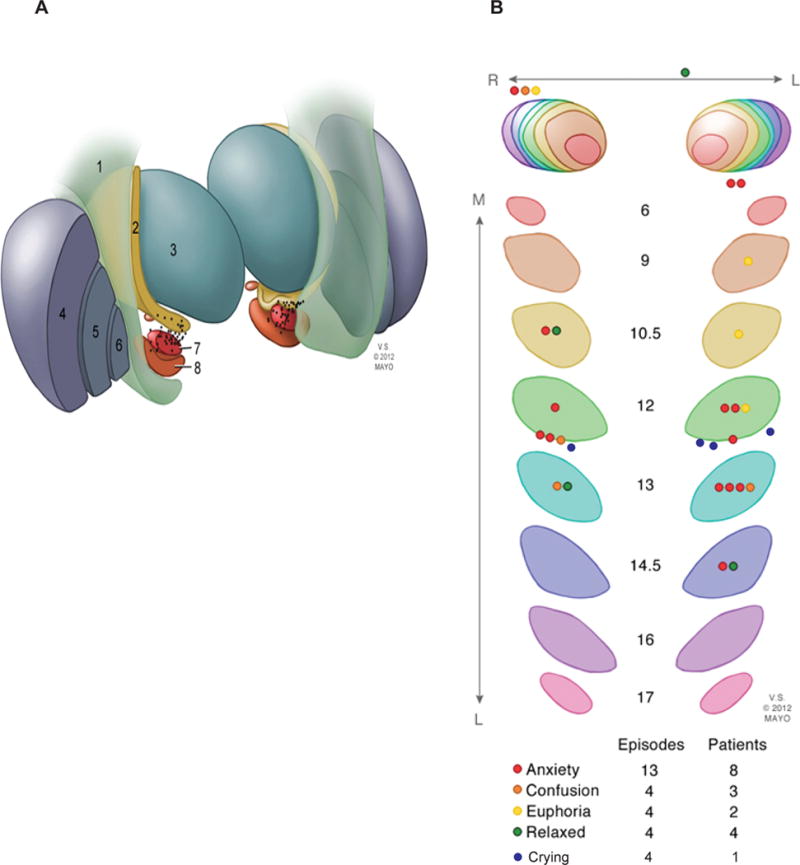
Figure-2. Neuroanatomical localization of active contacts.

The spatial location of the active contacts on postoperative MR images fused with the Schaltenbrand and Wahren human brain atlas. (A) Illustration demonstrating the anatomic location of the active contacts in relation to surrounding structures: 1. corticospinal tract, 2. zona incerta, 3. thalamus, 4. putamen, 5. globus pallidus external segment, 6. globus pallidus internal segment, 7. subthalamic nucleus, 8. substantia nigra. (B) Serial sections of the STN corresponding to slices in the Schaltenbrand and Wahren human brain atlas on the left (L6–L17) and right (R6–R17) showing the locations of active contacts and corresponding TNM categories.
