
Figure 3. Juxtaposition of WNThi and WNTlo Borders Is Sufficient to Acquire Asymmetric Cell Fates.
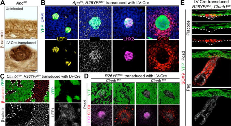
(A) Immunohistochemistry of Apcfl/fl epidermis uninfected (top) or LV-Cre-transduced (bottom), generating Apc-null regions with increased WNT activity, indicated by intense β-catenin signal.
(B) Planar confocal IMF of Apcfl/fl; R26YFPfl/+ epidermis mosaic for LV-Cre. Note WNT-hyperactivated next to WNT-normal regions establish boundary for asymmetric cell fates.
(C) Planar confocal IMF in Ctnnb1fl/fl; R26YFPfl/+ embryos transduced with LV-Cre. Note selective loss of LEF1 in IFE patches where β-catenin is absent.
(D) Planar confocal IMF in Ctnnb1fl/fl (or fl/+); R26YFPfl/+ embryos infected with high-titer LV-Cre to generate small regions of β-catenin+ untransduced cells surrounded by β-catenin− transduced cells.
(E) Sagittal IMF in placode (top) and peg (bottom) of R26YFPfl/+; Ctnnb1fl/fl embryos transduced with LV-Cre. Bracket indicates SOX9+ IFE halo. Note YFP+ SOX9+ IFE cells do not contribute to mature HF.
Tissues processed as indicated for immunohistochemistry (β-catenin) or IMF (LEF1, Pcad, LHX2, SOX9, β-catenin, YFP).
Dashed lines in (A,E) indicate basement membrane, in (C), borders between transduced and untransduced regions.
Scale bars in (A), 10μm; in (B,D,E), 20μm; in (C), 50μm.
See also Figure S2.