
Figure 7. SHH+ Cells Generate SOX9+ Stem Cells Early but Differentiation Lineages Late in HF Morphogenesis.
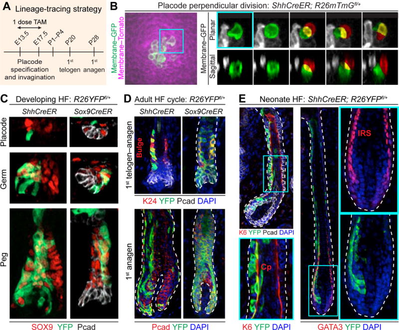
(A) Strategy to lineage-trace embryonic SHH+ and SOX9+ cells. ShhCreER; R26mTmGfl/+, ShhCreER; R26YFPfl/+, and Sox9CreER; R26YFPfl/+ embryos were treated with tamoxifen (TAM) once during window of HF morphogenesis and harvested at indicated times.
(B) Live imaging of perpendicular division of SHH+ placode cell. (Left) Labeled membrane shows SHH+ (GFP+) cells within a placode. (Right) Time course of maximum-intensity projections of planar confocal stacks (Top) and sagittal reconstructions (Bottom) of perpendicular division of SHH+ placode cell (green) leading to a basal (green) and suprabasal (red) cell (overlap, yellow). t=0 corresponds to onset of mitosis as determined by cell rounding. See Movie S5.
(C) Examples of lineage tracings of SHH+ cells marked at early stages of HF morphogenesis. Note that SHH+ cells give rise to SOX9+ cells, but SOX9+ cells do not generate Pcadhi SHH+ cells.
(D) Examples of lineage tracings monitored to 1st telogen and anagen. Note ShhCreER; R26YFP+ (and Sox9CreER) labeled cells contribute to adult SC pool (K24+) (bracket), and to all HF lineages in subsequent hair cycle (bottom).
(E) Examples of lineage tracings from cells marked at later stages of HF morphogenesis and monitored to HF maturation (P1–4). Note that SHH+ cells marked at later times give rise to differentiated lineages: K6, companion layer (Cp, arrows); GATA3, inner root sheath (IRS, arrows).
Tissues processed as indicated for IMF (SOX9, YFP, Pcad, K24, K6, GATA3) or endogenous fluorescence (GFP, Tomato).
White dashed lines indicate basement membrane.
All scale bars, 10μm.