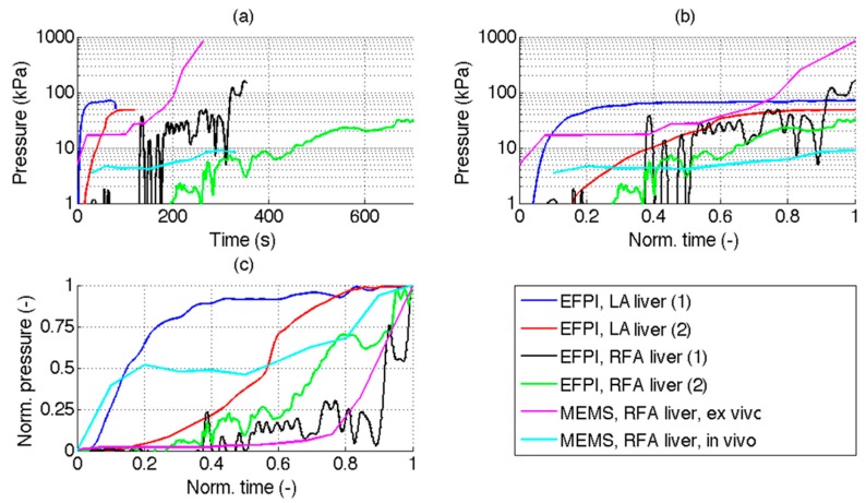
Figure 6.
Comparison between experimental results and previous pressure measurements in mini-invasive TA, documented in literature. The chart compares EFPI pressure measurement performed in LA on hepatic tissue, reporting experiments 1 and 2 from Figure 5; experimental results obtained with a similar EFPI probe by Tosi et al. [25] on RFA ablation of liver, at 0.1 cm (1) and 0.5 cm (2) distance between the probe and the applicator; experimental ex-vivo results of Kotoh et al. [16], obtained with a MEMS sensor at 3 cm distance between the ablation device and probe; in-vivo study by Kotoh et al. [17] for hepatic RFA on animals, recorded with a MEMS sensor. Data are reported in three different formats: (a) Pressure (logarithmic units) as a function of time; (b) Pressure (logarithmic units) and normalized time; (c) Both pressure and time are normalized. Normalized data are within [0, 1] range whereas 0 corresponds to ablation start and 1 corresponds to peak pressure, and its corresponding elapsed time. MEMS experiments have been digitized from [16] and [17].

