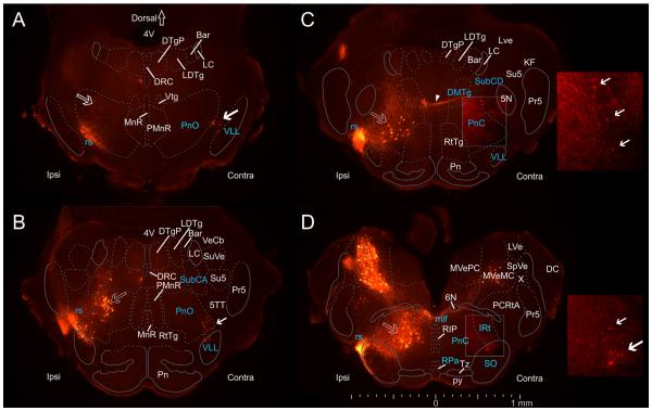
Figure 2.
Spatial distribution of pRS neurons and other neuron populations in the pons. Epifluorescence images (50-μm transverse sections, evenly spaced at 250-μm intervals through the pons at P0) showing retrograde labeling in the pons after applying RDA unilaterally (Ipsi) to the entire VF + LF at C2. Cytoarchitectonically defined boundaries of relevant nuclei and structures adapted from the atlas of Paxinos et al. (2007) have been superimposed (see Materials and Methods). Dashed lines indicate boundaries transferred from the Paxinos et al. (2007) atlas, which should thus be considered only as approximate, whereas solid lines indicate boundaries that could be seen in neighboring sections stained with methylene blue. A: Rostralmost section, showing ipsilateral pRS neurons in the PnO (open arrow) lying dorsal to the labeled axons of the rubrospinal tract (rs). Contralateral pRS neurons (solid arrow) lie near the lateral edge of the PnO, in the border area between the PnO and the ventral nucleus of the lateral lemniscus (VLL). B: At this level (250 μm more caudal than A), ipsilateral pRS neurons form a distinct cluster in the lateral half of the PnO, adjoining the more dorsal group of retrogradely labeled neurons in the nucleus subcoeruleus alpha part (SubCA). Contralateral pRS neurons lie in the ventrolateral corner of the PnO, similar to their location in A. C: Ipsilateral pRS neurons are located centrally in the PnC. A few contralateral pRS neurons are also labeled in the lateral part of the PnC. However, they are weakly labeled and are best seen in the inset displaying the same region at twice the magnification and with enhanced brightness and contrast (solid arrows indicate individual neurons). At this level, axons from contralateral pRS are seen traversing the PnC, the SubCD, and the dorsomedial tegmental area (DMTg) before crossing the midline (arrowhead). D: In the most caudal region of pons, ipsilateral pRS neurons lie centrally within the PnC where they are enmeshed with labeled axons coursing toward the spinal cord. Contralateral pRS neurons are located near the ventrolateral corner of the PnC, in a region that according to Paxinos et al. (2007) corresponds to the superior olive (SO) and the intermediate reticular nucleus (IRt). They are also shown in the inset at right. At this level, ipsilateral and contralateral vestibulospinal neurons can also be seen clearly. Note that the location of contralateral pRS neurons outside the indicated confines of the PnO is in our view due to a combination of shifts in the locations of boundaries from the illustrated section to the adjacent methylene blue-stained section and ambiguity in the transfer of the boundaries defined by Paxinos et al. (2007).

