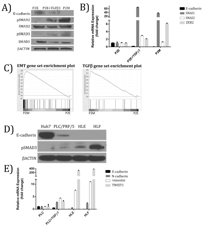
Figure 2. Active TGFβ signaling correlates with mesenchymal phenotype in murine and human models of HCC.
(A) Treatment of a mouse epithelial HCC cell line (P2E) with TGFβ at 5 ng/ml for 48 hours produces a molecular phenotype similar to mouse mesenchymal HCC cell line (P2M). (B) Up-regulation of mRNA levels of EMT markers in mouse epithelial HCC with TGFβ treatment (5 ng/ml for 48 hours). (C) Gene set enrichment analysis (GSEA) supports P2M cells are enriched for mesenchymal markers relative to P2E cells (left panel). GSEA reveals that TGFβ signaling is enriched in P2M versus P2E cells, suggesting that TGFβ may be driving the EMT phenotype in this mouse model of HCC (right panel). This gene set contains genes up-regulated by TGFβ1 in MCF10A cells. (FDR = 0.031). (D) Human mesenchymal HCC cell lines HLE and HLF have low E-cadherin expression and high pSMAD3, whereas epithelial-like HCC cell lines Huh7 and PLC/PRF/5 (PLC) express high E-cadherin and low pSMAD3. (E) Relative to the PLC/PRF/5 cell line, PLC/PRF/5 treated with TGFβ (10 ng/ml, 48 hrs), HLE, and HLF cell lines exhibited lower E-cadherin and higher vimentin, and TWIST1 expression. FDR = False discovery rate.

