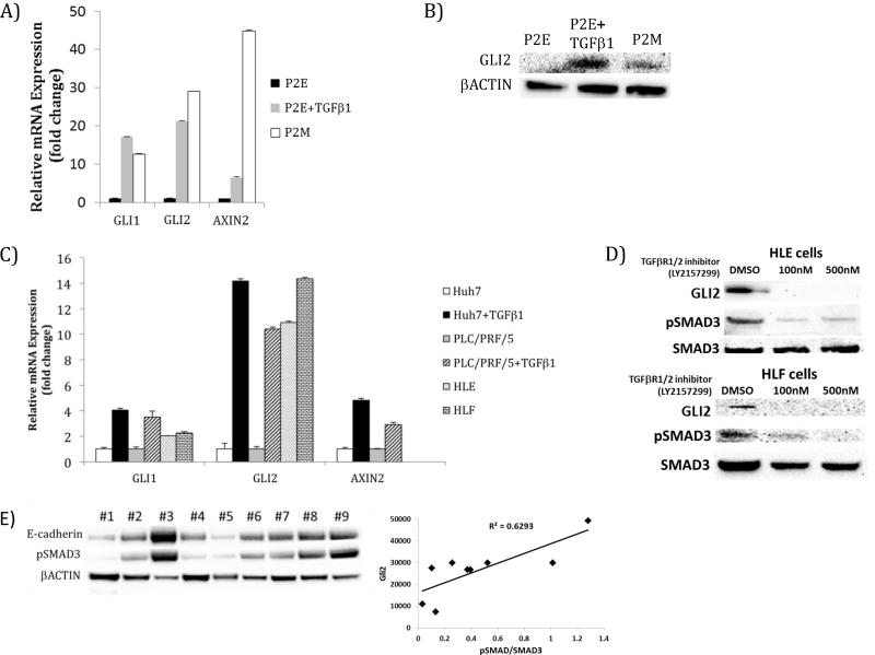
Figure 6. Network model-directed testing validates activation of Sonic hedgehog and Wnt signaling pathways by TGFβ.
A) GLI and AXIN2 transcripts are elevated in murine mesenchymal cell line P2M and by TGFβ treatment (5 ng/ml, 48 hours) of murine epithelial HCC cell line, P2E. B) GLI2 expression is elevated by TGFβ1 treatment (5 ng/ml, 48 hours) of epithelial P2E cells and in mesenchymal P2M cells relative to P2E cells. C) Human epithelial-like HCC cell line Huh7 has low expression of GLI1, GLI2, and AXIN2 and these transcripts are increased by TGFβ1 treatment (5 ng/ml, 48 hours). Epithelial-like PLC/PRF/5 cells treated with TGFβ1 (10 ng/ml, 48 hours) exhibit increased GLI1 and GLI2 expression. GLI1 and GLI2 expression were also elevated in mesenchymal phenotype cell lines with constitutively active TGFβ signaling, HLE and HLF. Treatment of PLC/PRF/5 cells with TGFβ1 (10 ng/ml, 48 hours) led to increased AXIN2 expression. D) Treatment of HLE and HLF cells with TGFβRI inhibitor, led to dose dependent loss of GLI2 levels. E) In protein extracted from nine human HCC patient biopsies, GLI2 expression correlated with pSMAD3 levels (R2= 0.6293).

