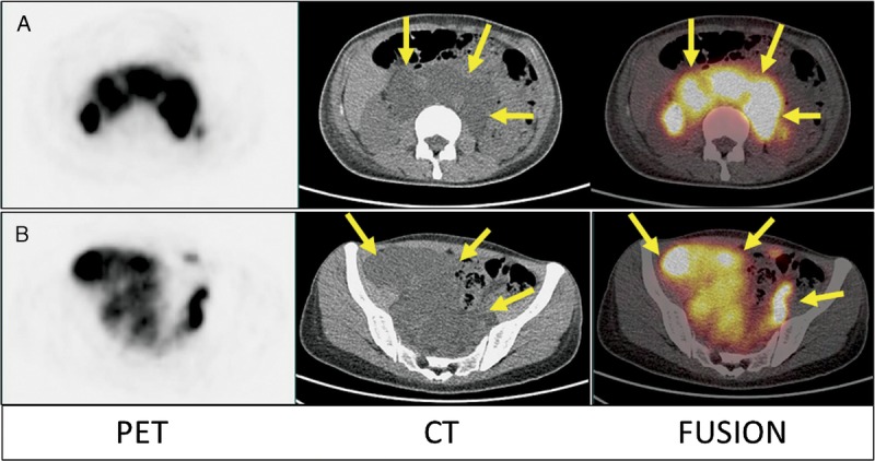
FIGURE 7.

Furthermore, the transaxial images of the abdomen (A) and pelvis (B) of 68Ga-NEB PET/CT from the same patient demonstrated that all of the hypodense cystic structures were filled with radioactive lymph fluid (small arrows), which was not impressively seen on 99mTc-SC scintigraphy. Eventually, the patient was proven to have lymphangioleiomyomatosis.
