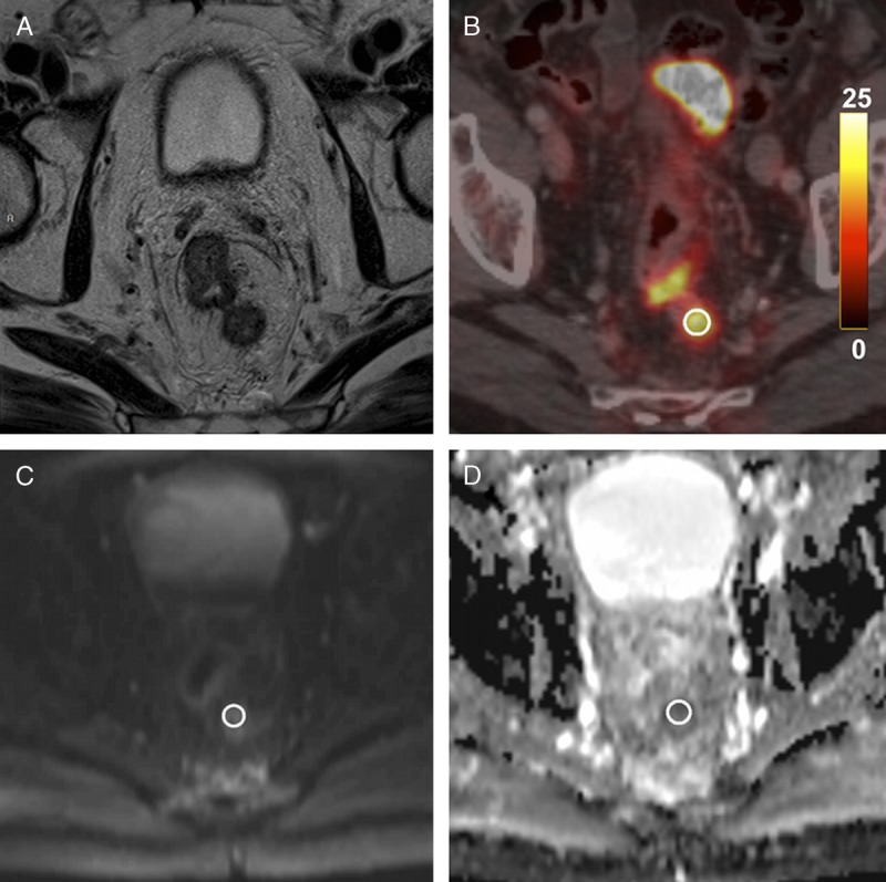
FIGURE 2.

Pathological lymph node in a 69-year-old man with a T4 N2 stage rectal cancer. A, Axial T2-weighted MR image shows a pathological lymph node located in the posterior mesorectum. B, Axial 18F-FDG PET/CT demonstrates high 18F-FDG uptake of this same lymph node (SUVmean = 8.9 g/mL, SUVmax = 10.7 g/mL). C and D, Axial diffusion-weighted image and ADC map show a focal diffusion restriction within the lymph node (ADCmean = 1047 × 10−6 mm2/s, ADCmin = 763 × 10−6 mm2/s).
