
Figure 3. m6A modification promotes selective translation under heat shock stress.
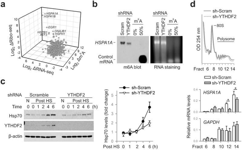
a, A 3-D plot depicting fold changes (log2) of mRNA abundance, CDS ribosome occupancy, and 5′UTR m6A levels in MEF cells after heat shock stress. b, m6A blotting of HSPA1A purified from MEF with or without YTHDF2 knockdown. mRNAs synthesized by in vitro transcription in the absence or presence of m6A were used as control. Representative of two biological replicates. c, Immunoblotting of MEF cells with or without YTHDF2 knockdown after heat shock stress (42°C, 1 h). N: no heat shock. The right panel shows the relative protein levels quantified by densitometry and normalized to β-actin. Representative of three biological replicates. d, MEF cells with or without YTHDF2 knockdown were subject to heat shock stress followed by sucrose gradient sedimentation. Specific mRNA levels in polysome fractions were measured by qPCR. The values are first normalized to the spike in control then to the total. Error bars, mean ± s.e.m.; * p < 0.05, unpaired two-tailed t-test; n=3, biological replicates (c and d).