
Ext. Data Fig. 3. C-Myc Expression becomes asymmetric in late telophase.
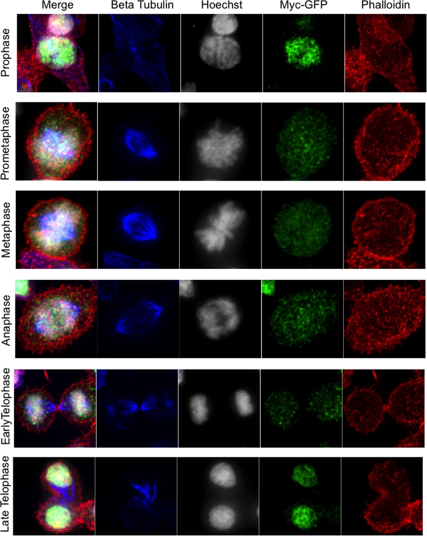
Overlay, Beta Tubulin (blue), Hoechst 33258 (gray), c-Myc-GFP (green), and phalloidin (red) for each mitotic phase indicated on the left as identified by chromatin and tubulin staining patterns.