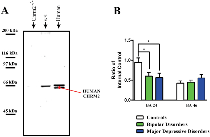
Figure 2.
(A) A Western blot showing immunogenic bands visualised in cortex from muscarinic M2 receptor knock out mouse (Chrm2-/-), wild type mouse (w/t) and human cortex (human) with the anti-muscarinic M2 receptor antibody used to quantify muscarinic M2 receptor protein in human cortex.
(B) Levels (mean ± SEM) of muscarinic M2 receptors in Brodmann’s areas (BA) 24 and 46 from subjects with bipolar disorder, major depressive disorder and age and sex matched controls.
*p < 0.05

