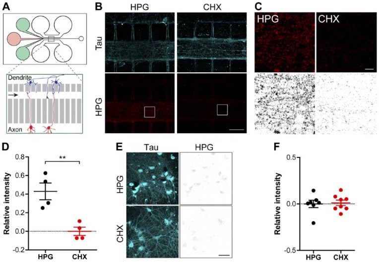
Figure 4.
Imaging of protein synthesis in neurons grown in microfluidic perfusion chambers. (A) Schematic overview of a microfluidic perfusion chamber with one region (green square) enlarged (inset of the perfusion channel on the right). On the left side of the chamber, there are three reservoirs. The two outer green wells contain a buffer to hold the perfusate inside the perfusion chamber preventing it from entering the microgrooves. The middle red well contains the desired perfusate. In the enlargement image, the red cells (lower) generate the axons growing towards the perfusion channel (presynaptic compartment), while the blue cells (upper) generate dendrites (postsynaptic compartment). (B) Representative confocal images of the HPG signal (red and greyscale) and Tau (blue) within the perfusion channel of chambers in which mature cortical neurons (DIV 14–16) were cultured. The neurons were treated locally within the perfusion channel with HPG alone or in combination with CHX. (C) Magnifications of the white square inserts depicted in (B) with the HPG shown in red and inverted greyscale. (D) Quantification of the relative intensity of the HPG signal inside the perfusion channel compared with neurons treated with CHX. (E) Representative confocal images of cells on the dendrite side (postsynaptic compartment) of the perfusion chamber with Tau (blue) and the HPG signal (greyscale). (F) Quantification of the relative intensity of the HPG signal of cells cultured on the dendrite side of the perfusion chamber. Data represent the mean ± SEM and single values shown for n=4 independent experiments. p-values are determined by two-tailed unpaired Students t-test. p<0.01. CHX, cyclohexamide; DIV, days in vitro; HPG, homopropargylglycine. **Scale (B and E) 50 µm; (C) 5 µm.

