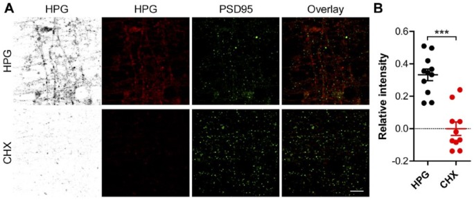
Figure 5.
Visualization of nascent protein synthesis in postsynaptic densities from neurons grown in microfluidic perfusion chambers. (A) Representative confocal images of the perfusion channel from microfluidic perfusion chambers in which DIV 14-16 primary cortical neurons were grown. Neurons were treated with HPG alone or in combination with CHX and stained for HPG incorporation (inverted greyscale and red) or PSD-95 (green), the overlay shows the overlap between these two signals. (B) Quantified relative HPG signals inside PSD-95 puncta of neurons treated HPG alone or in which translation was inhibited using CHX, normalized for the total selected PSD-95-positive-area. Data represent the mean ± SEM and single values shown for n=10 to 11 culture chambers collected from three independent experiments. CHX, cyclohexamide; DIV, days in vitro; HPG, homopropargylglycine; PSD-95, postsynaptic density protein 95. p-values are determined by two-tailed unpaired Students t-test. ***p<0.0001. Scale, 5 µm.

