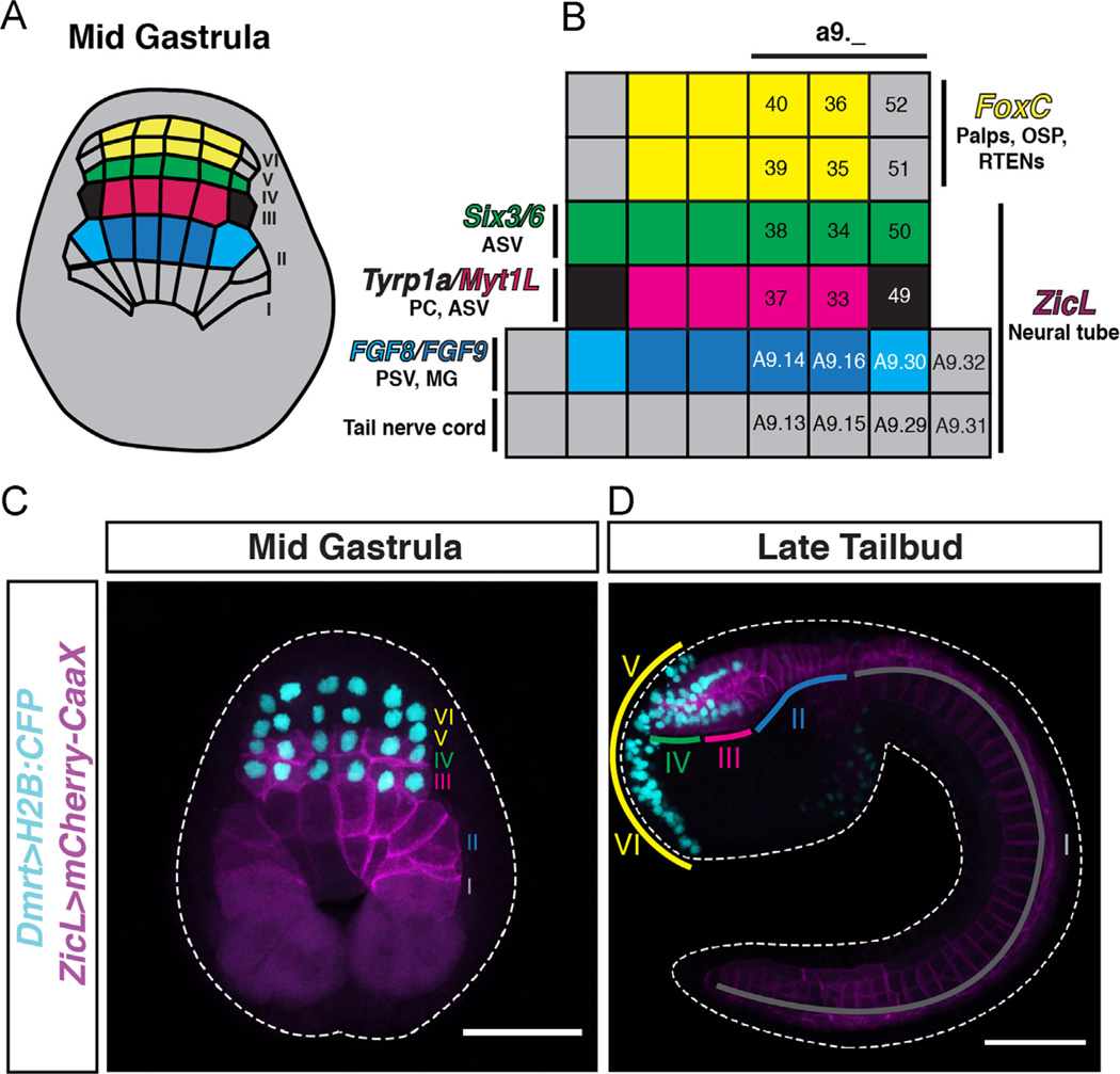
Fig. 1.
The Ciona mid-gastrula neural plate. (A) Schematic of a mid-gastrula stage embryo showing the organization of the 6-row neural plate. (B) Gene expression patterns and fates of neural plate territories. Rows V and VI express FoxC and give rise to the adhesive palps, oral siphon placode (OSP) and rostral trunk epidermal neurons (RTENs). Row IV expresses Six3/6 and contributes to the anterior sensory vesicle (ASV). The a9.49 cells in lateral row III express Tyrp1a and give rise to pigmented cells (PC). Medial row III cells (a9.33 and a9.37) express Myt1L and also contribute to the ASV. The FGF8-expressing A9.30 cells in lateral row II form the motor ganglion (MG), whereas medial row II forms posterior sensory vesicle (PSV). Row I contributes to the tail nerve cord. (C, D) Co-electroporation of Dmrt and ZicL reporters allows for tracing of cells from the mid-gastrula neural plate (C) to the late tailbud stage (D). Colored lines in (D) indicate territories derived from rows labeled with the corresponding color in (C). Rows V and VI express Dmrt exclusively and contribute to the palps, OSP, and peripheral nervous system. ZicL and Dmrt reporters co-localize in rows III and IV, which give rise to the anterior sensory vesicle. Rows I and II express ZicL exclusively and contribute to posterior brain, motor ganglion, and tail nerve cord. Scale bars in (C, D) = 50 µm.

