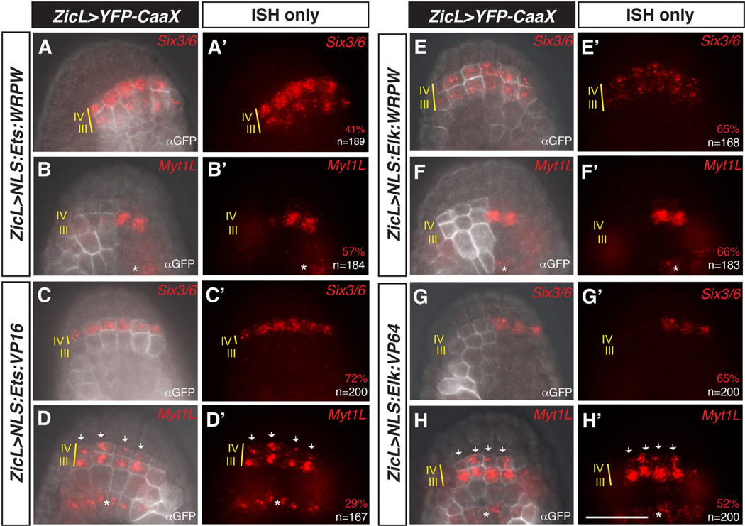
Fig. 3.
Partially redundant roles for Ets1/2 and Elk1/3/4 in the anterior neural plate. (A–D′) Embryos were co-electroporated with ZicL > YFP-CaaX and ZicL > Ets:WRPW (A–B′) or ZicL > Ets:VP16 (C–D′), and then assayed for expression of Six3/6 (A, A′, C, C′), or Myt1L (B, B′, D, D′) at mid-gastrula stage by in situ hybridization and immunohistochemistry with GFP antibody (αGFP). ZicL > Ets:WRPW leads to modest expansion of Six3/6 (A, A′) and loss of Myt1L (B, B′). ZicL > Ets:VP16 embryos show mostly wild-type expression of Six3/6 (C, C′) and a moderate expansion of Myt1L (arrows in D, D′). (E–H′) Similar to (A–D′), except embryos were electroporated with ZicL > Elk:WRPW (E–F′) or ZicL > Elk:VP64 (G–H′). ZicL > Elk:WRPW has a slightly stronger effect on Six3/6 expansion into row III (E, E′). This expansion occurs at the expense of Myt1L (F, F′). ZicL > Elk:VP64 results in repression of Six3/6 in row IV (G, G′) and expansion of Myt1L expression (arrows in H, H′). Embryos in (B, B′), (F, F′) and (G, G′) are left–right mosaics, owing to unequal inheritance of electroporated plasmids. Scale bar = 50 µm. Asterisks indicate expression of Myt1L in row I. See full quantitation of observed phenotypes in Supplementary Fig. 5.

