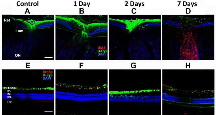
Fig 1. Intraocular distribution of intravitreally injected Cy5-labeled dendrimers (D-Cy5, green) in rNAION and control eyes.
60μg of D-Cy5 were injected intravitreally into either control or rNAION-induced eyes as described in the Methods section and Table 1. Animals were euthanized, and the posterior portion of the eye, including retina and first portion of the optic nerve (A-D) were examined, along with isolated retina (E-H), and analyzed for dendrimer uptake (in green) at 1, 2 or 7 days post-injection. Minimal signal is present in the laminar and retrolaminar ON in either control or rNAION-induced eyes at any time point; however, there is marked signal in the optic disc and inner layers of the peripapillary retina in the control eye and at both 1- and 2- days in the rNAION-induced eyes. By 7 days post-injection, there is only faint signal in the optic disc and peripapillary retina of the rNAION-induced eyes. In all panels, Green = D-Cy5 and Blue = DAPI. Red varies with figure as noted on the figure. A-D: Red = IBA1. E-H: Red = Brn3a. Scale bar (optic nerve): 200μm. Scale bar (retina): 100μm.

