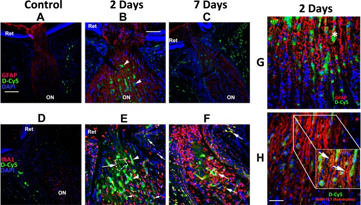
Fig 2. Fluorescent dendrimer distribution in the laminar region and distal optic nerve following rNAION induction and intravenous injection of Cy5-labeled dendrimers (D-Cy5).
Cy5-labeled dendrimers were administered intravenously to animals in which rNAION was induced in one eye as indicated in the Methods section and Table 2. Animals were euthanized and optic nerves (ON) analyzed for dendrimer uptake (in green) in control animals and in rNAION animals 2 or 7 days post-induction. Confocal analysis reveals no accumulation of dendrimer in retinas or ONs of non-induced eyes at any timepoint, except in the ON sheath. Substantial dendrimer signal is seen in the ischemic region in rNAION-induced eyes at 2 days (B,E) and 7 days (C,F). G and H: Confocal analysis of astrocyte-associated dendrimer uptake at 2 days post-induction. G: GFAP (astrocyte structural protein)/Cy5 dendrimer colocalization. Dendrimer signal is found in both swollen sac-like structures associated with capillaries (asterisk) and in a linear pattern (arrows in B). H: Aldh1L1 (astrocyte-specific cytoplasmic protein)/dendrimer co-localization. There is extensive dendrimer signal overlap with the astrocyte cytoplasm (arrows in inset in H). There is also slight signal in ON sheaths of both non-induced and rNAION-induced eyes, presumably in macrophages. A-C and G, Red = GFAP. D-F, Red = IBA1. H, Red = Aldh1L1. Scale bars: A,C,D: 200μm. B,E,F: 100μm. Scale bar in G,H: 50μm. Scale bar in inset: 20μm.

