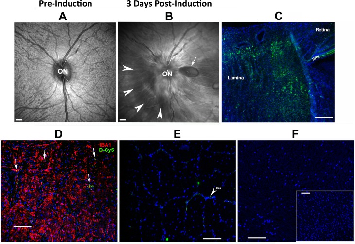
Fig 5. D-Cy5 is present in the region of optic nerve ischemia in eyes with pNAION but not in white or gray matter in the brain.
A. SD-OCT baseline scan of the primate optic disk and surrounding retina (en face view). The optic disk (ON) and retinal nerve fiber layer entering the nerve are flat against the back of the eye. The underlying choroidal vasculature is visible through the retina. B. SD-OCT en face scan of the posterior portion of the same eye 3 days post-pNAION induction. The disk (ON) is grossly swollen, with blurring of the disk margin. There is a serous peripapillary retinal detachment (arrowheads), and a peripapillary retinal nerve fiber layer hemorrhage (arrow). C-F: Histology post-pNAION of the same animal 3d post-D-Cy5 injection (6d post-induction). C. Laminar region, longitudinal section. D-Cy5 is concentrated in the laminar region to a depth of 1mm (approximately the entire laminar thickness). D-Cy5 is also present in the RPE. D. Lamina, IBA1 immunohistochemistry. D-Cy5 co-localizes in individual laminar inflammatory cells (arrows) that are concentrated in the pNAION lesion. E. Distal ON. There is minimal D-Cy5 signal that is restricted to small areas of optic nerve connective-tissue septae (sep; arrowhead). F. CNS white and gray matter. There is minimal D-Cy5 signal in both white (corpus callosum) and gray (inset: cortex) matter. Scale bars in A and B: 500μm, Scale bar in C: 200μm. Scale bars in D,E and F: 100μm. Scale bar in F (inset): 100μm.

