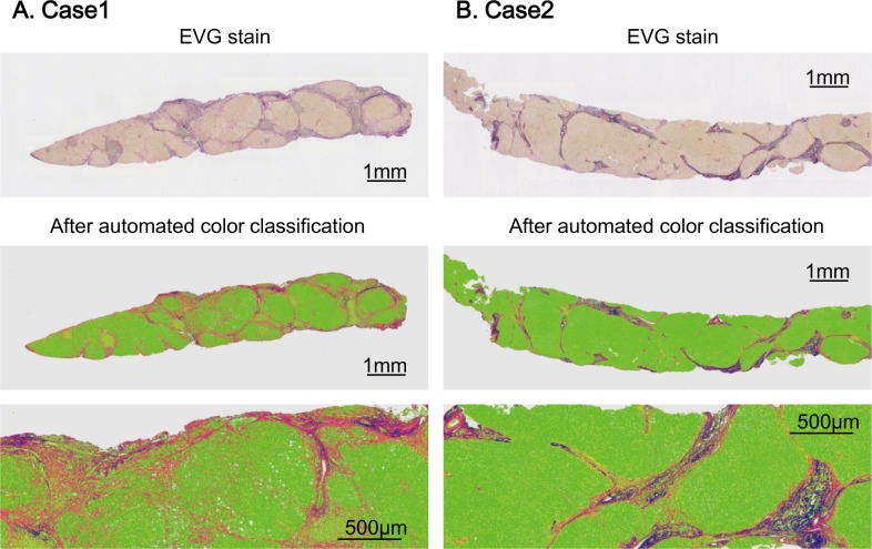
Fig 1. Representative cases of fiber quantification.
Panel (A) and (B) show representative cases of automated fiber quantification. Both cases were diagnosed as F4 by METAVIR staging system. After automated color classification, collagen fiber was depicted as red color whereas elastin fiber was depicted as blue color. Case 1 is a patient with relatively low elastin proportional area, who showed 14.1% of collagen and 2.5% of elastin. On the contrary, case 2 is a patient with a higher elastin, who showed 6.5% of collagen and 4.8% of elastin.

