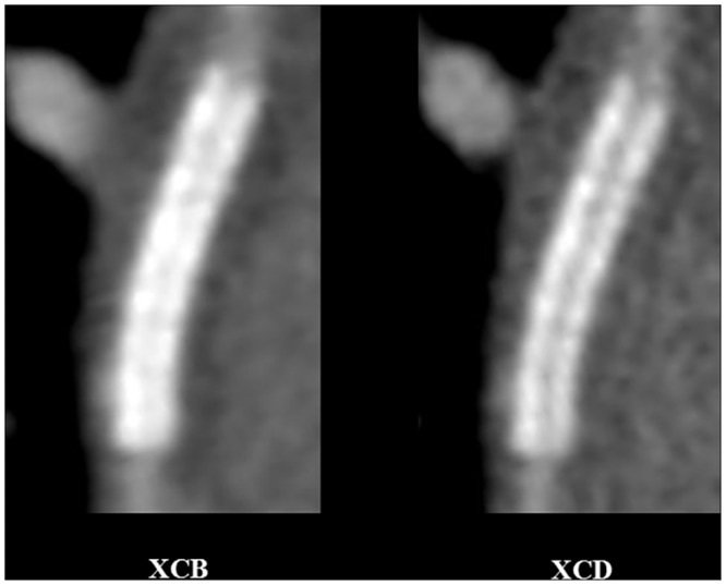
Fig 5. Reduced stent blooming artifacts and improved strut definition with sharp (XCD) in comparison to smooth (XCB) kernel.

69-year-old female, first obtuse marginal artery stent. 256-slice CT acquisition with prospective ECG-gating, and image reconstuction with a medium-soft (XCB, left) and edge-enhancing (XCD, right) reconstruction kernels, multiplanar reformat. Window width, 1500 HU; window centre: 300 HU, for both kernels. For observer 1, stent wall thickness for the XCB and XCD kernels with the orthogonal thickness method was 1.29 mm and 1.05 mm, and 1.26 mm and 1.26 mm with the circumference method, respectively. Image quality scores for observer 1 were 3 and 4, respectively. For observer 2, stent wall thickness for the XCB and XCD kernels with the orthogonal thickness method was 0.97 mm and 0.71 mm, and 0.82 mm and 0.83 mm with the circumference method, respectively. Image quality scores for observer 2 were 2 and 3, respectively.
