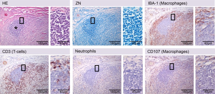
Fig 1. Established immunohistochemical stainings for leukocytes in the pig skin.
Serial histologic sections of pig skin stained with Haematoxylin/Eosin (HE), Ziehl-Neelsen/Methyleneblue (ZN) and by immunohistochemistry protocols established for antibodies specific for T-cells (CD3), macrophages (IBA-1, CD107a) or neutrophils (21 kDa neutrophil protein). A B-cell cluster is shown that can only be identified by exclusion criteria (asterisk). The cluster of cells (asterisk) was not stained positive with antibodies against T-cells (CD3), macrophages (IBA-1, CD107a) nor neutrophils (Neutrophils), while the cells have lymphocyte appearance (ZN, insert).

