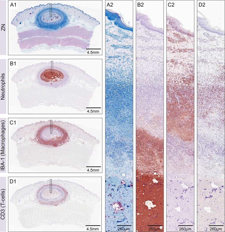
Fig 2. Layers of leukocyte infiltration in nodular lesions six weeks after infection.
Histological sections of a nodular lesion six weeks after infection with 1.3 x 10⁶ CFU of M. ulcerans. Ziehl-Neelsen/Methyleneblue (ZN) staining revealed a strong cellular infiltration around a central necrotic core containing AFB stained in pink (A1, A2). The necrotic core and the surrounding ring of infiltration consisted of neutrophils (B1, B2). Neutrophils were surrounded by a belt of macrophages (C1, C2) that were heavily interspersed with T-cells (D1, D2).

