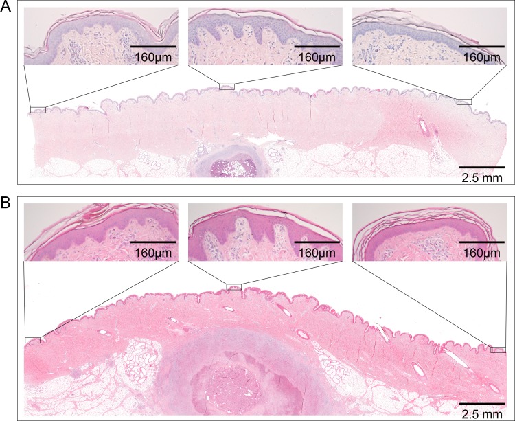
Fig 3. Epidermal hyperplasia above nodular lesions.
Histological sections of nodular lesions stained with Haematoxylin/Eosin. (A) Small nodule caused by infection with 1.3 x 10⁵ CFU M. ulcerans. (B) Large nodule caused by infection with 1.3 x 10⁶ CFU M. ulcerans. Epidermal hyperplasia was strongest directly above the lesion (central box) and weaker if further away from the lesion.

