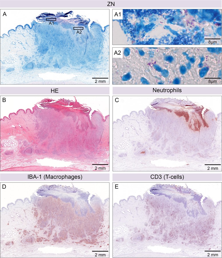
Fig 4. Ulcerative lesion after six weeks of infection.
Histological sections of an ulcerative lesion six weeks after infection with 1.3 x 10⁶ CFU M. ulcerans. Ziehl-Neelsen/Methyleneblue (ZN) staining revealed AFB and bacterial debris in the crust (pink, A1) together with secondary infection (cocci, small blue dots, A1). More AFB were found just below the opening of the ulcer (A2). Haematoxylin-Eosin staining of the whole lesion (B). Neutrophils were predominantly located just below the opening of the ulceration (C) and in an additional focus in the subcutaneous fat layer. The majority of cellular infiltration consisted of macrophages (D) interspersed with a high number of T-cells (E).

