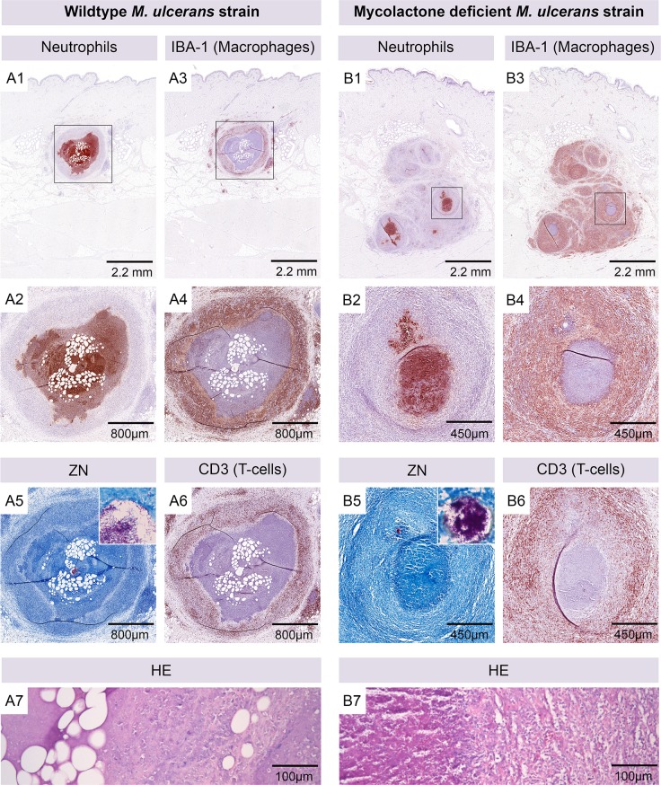
Fig 5. Different histopathological appearance of lesions caused by mycolactone producing and non-producing M. ulcerans strains.
Histological sections of a nodular lesion six weeks after infection with 1.3 x 10⁵ CFU wild-type M. ulcerans (A). The organisation of cellular infiltration around a necrotic core containing AFB (A5) and fat cell ghosts (A7) was comparable to lesions caused by higher doses of M. ulcerans. The central part of the nodule contained neutrophils (A1, A2), surrounded by a ring of macrophages (A3, A4) that was interspersed with T-cells (A6). Infection with 3.1 x 10⁵ CFU of mycolactone-deficient M. ulcerans led to the development of a lesion with more inhomogeneously organized infiltration (B). Several granulomatous structures contained small cores consisting of neutrophils (B1, B2) and AFB (B5). Macrophage infiltration was massive (B4) and interspersed with numerous T-cells (B6). Fat cell ghosts were absent and necrosis was less extensive (B7).

