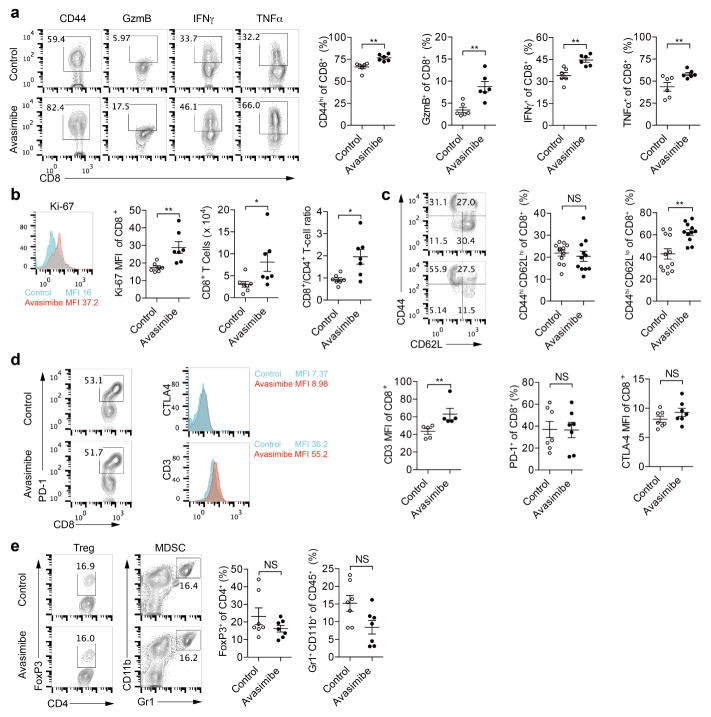
Extended Data Figure 10. Avasimibe treatment leads to potentiated effector function and enhanced proliferation of tumour-infiltrating CD8+ T cells.
C57BL/6 mice bearing B16F10 melanoma were treated with avasimibe (15 mg kg−1) or same dose of DMSO in PBS for four times by intraperitoneal injection. The mice were euthanized at day 18 and the tumours were isolated. a, The CD44 surface expression and cytokine/ granule productions of tumour-infiltrating CD8+ T cells were assessed using flow cytometry (n = 6). b, CD8+ T-cell number, CD8+/CD4+ T-cell ratio and Ki-67 level of tumour-infiltrating CD8+ T cells were assessed using flow cytometry (n = 7). c, Percentages of CD8+ central memory (CD44hiCD62Lhi) and CD8+ effector/effectory memory (CD44hi CD62Llo) were assessed using flow cytometry (control, n = 12; avasimibe, n = 11). d, Surface levels of PD-1, CTLA-4 and TCR of tumour-infiltrating CD8+ T cells. PD-1 level was indicated by the percentage of PD-1hi cells. CTLA-4 and TCR levels were indicated by median fluorescence intensity. PD-1 and CLTA-4 staining, n = 7; TCR staining, n = 5. e, Treg cell (FoxP3+ CD4+) and myeloid-derived suppressor cell (MDSC) (Gr1+ CD11b+) percentages in the tumour microenvironment were assessed using flow cytometry (n = 7). Data were analysed by Mann–Whitney test. Error bars denote s.e.m; *P < 0.05; **P < 0.01; ***P < 0.001.

