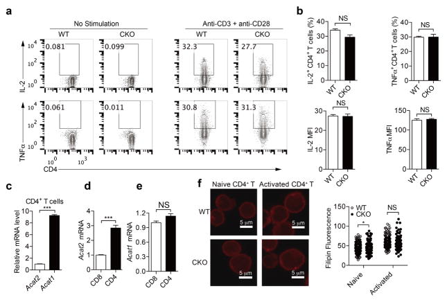
Extended Data Figure 4. ACAT1 deficiency does not result in significant change of CD4+ T-cell function.
a, b, Cytokine productions of CD4+ T cells (n = 3). Cells were stimulated with 5 μg ml−1 plate-bound anti-CD3 and anti-CD28 antibodies for 12 h. Representative flow cytometric profiles are shown in a. c–e, Relative transcription levels of Acat1 and Acat2 in naive CD4+ and CD8+ T cells freshly isolated from C57BL/6 mice (n = 3). Acat1 transcription level was significantly higher than Acat2 in CD4+ T cells. Acat1 transcription levels were comparable between CD4+ and CD8+ T cells, whereas the Acat2 transcription level in CD4+ T cells was significantly higher than that in CD8+ T cells. Acat2 transcription level in CD4+ T cells was set as 1 in c. Acat1 and Acat2 transcription levels in CD8+ T cells were set as 1 in d and e. f, Filipin III staining to analyse cellular cholesterol distribution in naive and activated CD4+ T cells from wild-type and CKO mice. Data were analysed by unpaired t-test (b–e) or Mann–Whitney test (f). Error bars denote s.e.m; *P < 0.05; ***P < 0.001.

