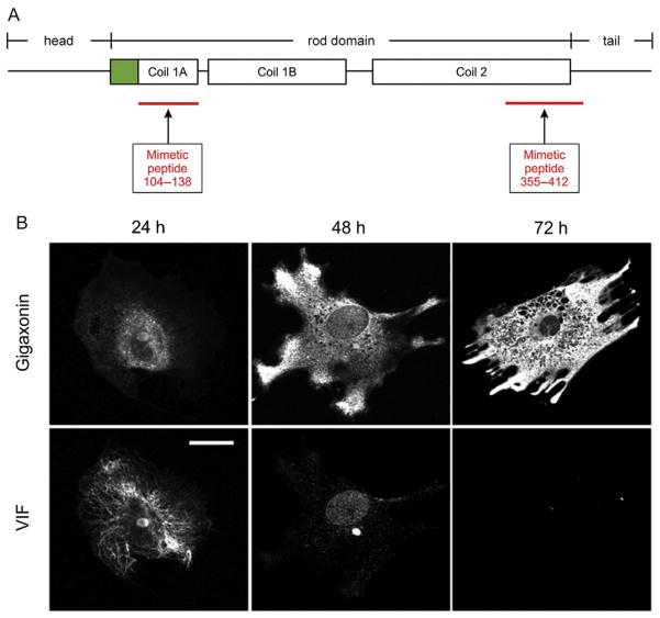
Figure 1.
Use of mimetic peptides and gigaxonin to study the cell biology of vimentin intermediate filaments (IFs). (A) Location of the mimetic peptides relative to the organization of a vimentin monomer. The monomer is divided into three major domains: head, α-helical rod, and tail. The rod domain is further segmented into coil 1 and coil 2. The diagram shows the secondary segmentation of coil 1 into 1A and 1B. Mimetic peptide 104–138 spans almost the entire coil 1A region. Mimetic peptide 355–412 includes part of the carboxy-terminus of coil 2 and seven amino acids of the tail. Green (gray in the print version) indicates the precoil domain. (B) Gigaxonin as an efficient tool for reducing and eliminating vimentin IFs from cells. Fibroblasts derived from a patient with giant axonal neuropathy were transfected with a mammalian expression vector-containing FLAG-tagged wild-type gigaxonin, fixed at the indicated times, and double-labeled with anti-FLAG (top row) and antivimentin (bottom row) antibodies. Twenty-four hours after induction of gigaxonin expression, vimentin IFs (VIF) were still observed. By 48 h, only short filaments and nonfilamentous vimentin particles remained; and by 72 h, there was no detectable vimentin. Scale bar, 10 μm. Images in (B) adapted from Mahammad et al. (2013).

