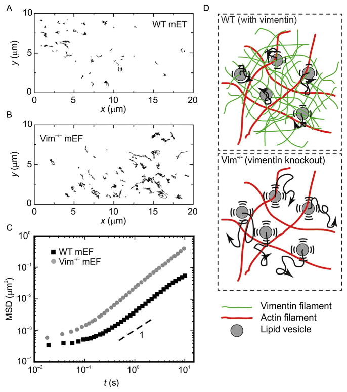
Figure 8.
Intracellular movement of endogenous vesicles and protein complexes inside wild-type (WT) and Vimentin−/− (Vim−/−) mouse embryonic fibroblasts (mEFs). (A, B) Ten-second trajectories of endogenous vesicles and protein complexes in the cytoplasm of (A) WT mEFs and (B) Vim−/− mEFs. These refractive objects are visualized by bright-field microscopy. (C) Calculation of the mean squared displacement of vesicles and protein complexes shows that these organelles move faster in the Vim−/− mEFs than in the WT mEFs. (D) Illustration of random organelle movement in networks with and without vimentin. In the WT cells, the vimentin network constrains the diffusive-like movement of organelles; in the Vim−/− cells, organelles move more freely.

